

For healthcare managers, access to imaging is not only a clinical concern. It also affects patient flow, room availability, staff workload and the speed of decision-making. A clinician may need a focused image at the bedside while the main ultrasound unit is being used elsewhere. Moving patients between departments can also create delays, especially in emergency and critical care.
These challenges are increasing interest in point-of-care ultrasound, or POCUS. It allows trained clinicians to perform focused examinations closer to the patient and can support faster assessments when introduced through clear clinical protocols.
Why Faster Access to Imaging Matters
Imaging delays can keep rooms occupied, increase transfer coordination and postpone clinical decisions.
Point-of-care ultrasound can help answer specific questions during the initial assessment. It may support procedural guidance, bedside monitoring and decisions about whether further diagnostic imaging or specialist referral is required.
Handheld devices should not, however, be presented as a complete replacement for radiology departments or comprehensive ultrasound examinations. Their main role is focused imaging performed by trained users for clearly defined clinical purposes.
What Handheld Ultrasound Brings to the Point of Care
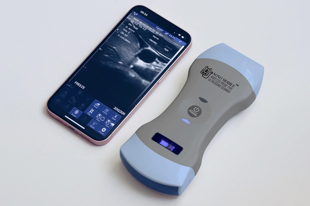
A modern Handheld Ultrasound can connect wirelessly to a compatible smartphone, tablet or computer and display real-time images at the bedside. Its compact design makes it suitable for examination rooms, emergency departments, procedure areas, intensive-care units and mobile medical services.
For managers, portability creates operational flexibility. The device can be assigned to one department, shared among selected teams or moved between rooms without the logistical demands of a cart-based system. This is especially useful in clinics where space is limited or focused ultrasound is needed only for certain patients and procedures.
Its value still depends on correct use. Facilities should confirm regulatory status, define approved applications and ensure that examinations are performed by appropriately trained professionals.
Operational Benefits for Healthcare Facilities
One of the main advantages of handheld ultrasound is improved equipment availability. Instead of waiting for a full-size system, clinicians may be able to perform an appropriate focused examination during the patient encounter.
Bedside access can also reduce unnecessary patient movement. This is valuable for patients with limited mobility, isolation requirements or unstable conditions. Even when further imaging is needed, an early focused assessment may help the team determine urgency and plan the next step.
Compact devices also allow smaller clinics to introduce selected ultrasound-guided services without dedicating an entire room to imaging equipment.
Where Handheld Ultrasound Can Add Value
The correct device depends on the center’s specialties. Different probes are designed for different imaging depths and anatomical structures.
A linear probe is commonly used for superficial anatomy, vascular access, musculoskeletal imaging and guided injections. A convex probe is more suitable for abdominal, renal and many obstetric examinations. Cardiac assessments require a suitable cardiac configuration, while women’s health and selected urology services may need convex or endocavitary options.
The most advanced model is not automatically the best purchase. A pain-management clinic may prioritize linear imaging and needle visualisation, while an urgent-care center may need a versatile configuration for abdominal, vascular and cardiac assessments. Purchasing should begin with real clinical use cases.
What Managers Should Evaluate Before Purchasing
Before approving a purchase, healthcare managers should review the device from clinical, operational and financial perspectives.
- Clinical suitability: The probe, imaging depth, presets and measurement tools should match the examinations and procedures the center intends to perform.
- Imaging capabilities: Depending on the application, the facility may need B-mode, M-mode, color Doppler, power Doppler or pulsed-wave Doppler.
- Compatibility and integration: Managers should confirm compatibility with existing phones, tablets and computers, as well as image storage, reporting, DICOM, PACS and electronic medical record requirements.
- Infection control: The center needs clear procedures for cleaning, disinfection, probe covers, storage and shared use.
- Training and quality assurance: Approved users, competency requirements, documentation standards and image review should be defined before routine use.
- Warranty and support: Repair procedures, replacement options, software support and service times can directly affect clinical downtime.
Looking Beyond the Initial Price
The purchase price is only one part of the total cost. Managers should determine whether the software requires subscriptions, whether important modes involve additional fees and which accessories are included.
Other costs may include tablets, protective cases, probe covers, training, integration and replacement equipment during repairs. A multi-year comparison is more useful than comparing initial prices alone.
Why Sono Mobile Is a Practical Option
Fagonex offers Sono Mobile in several configurations, allowing healthcare centers to choose a model according to their actual services.
The Sono Mobile L4 is designed for linear applications such as vascular access and musculoskeletal imaging. The C6 focuses on convex imaging for abdominal and general examinations. The CL64 combines convex, linear and cardiac capabilities for facilities requiring greater flexibility. The CT61 supports convex and vaginal applications for women’s health, obstetrics and selected urology services.
This range helps managers choose a configuration that matches the center’s procedures and clinical priorities.
The Sono Mobile application is available for compatible iOS, Android and Windows devices. Fagonex states that its software supports image management, reporting, DICOM, PACS and telemedicine without ongoing membership or subscription fees.
The absence of recurring software fees may help control long-term costs. Still, compatibility, cybersecurity, storage and integration should be tested within the center’s IT environment before full deployment.
A live demonstration lets clinicians assess image quality, IT teams review connectivity and managers compare models against the facility’s workflow.
Implementing the Device Successfully
Purchasing the device should be followed by a structured implementation plan. The center can begin with one department, a limited number of approved examinations and a small group of trained users.
Before routine use, the organization should define cleaning, documentation, storage and quality-review procedures. During the initial phase, managers can monitor device use, technical issues, training needs and the effect on patient movement and referrals.
A More Flexible Approach to Clinical Imaging
Handheld ultrasound can bring focused imaging closer to the patient and give healthcare facilities more flexibility in how they use rooms, staff and equipment. Its value depends on selecting the correct probe, training users and integrating the device into safe clinical processes.
Healthcare managers should compare models according to their actual services, regulatory requirements and total cost of ownership. Fagonex provides demonstrations and model-selection support for centers that want to assess how Sono Mobile can fit into their workflow before making a purchasing decision.